Exosome Isolation & Purification Services
OverviewServiceSamplesAdvantagesCase StudyFAQs
Overview
Microbial extracellular vesicles (mEVs) represent an emerging class of natural nanoparticles with significant potential in interspecies communication, immune modulation, and therapeutic delivery. Unlike mammalian exosomes, microbial EVs are secreted into complex fermentation media containing high concentrations of proteins, polysaccharides, metabolites, and cellular debris. Their isolation demands specialized workflows that account for small particle size (20–300 nm), cell wall complexity, and endotoxin contamination risks.
At Creative BioMart Microbe, we have developed a multi-platform purification infrastructure specifically optimized for microbial culture supernatants. Our scientists select and configure isolation strategies based on strain-specific secretion profiles, culture viscosity, and downstream analytical or functional requirements. Whether you require a small batch for NTA and TEM characterization, or a purified master bank for drug loading and in vivo efficacy studies, we provide reproducible, well-documented mEV preparations. This platform is a core downstream module of the Microbial Exosome Services suite.

Figure 1. Overview of Creative BioMart Microbe's multi-platform microbial extracellular vesicle (mEV) isolation and purification infrastructure. (AI-generated)
Our Service
Service Workflow
Our standardized workflow ensures traceability and reproducibility from sample receipt to final delivery.

Service Details
Differential Centrifugation & Ultracentrifugation
We perform sequential differential centrifugation to remove cellular debris and apoptotic bodies, followed by high-speed ultracentrifugation to pellet EVs. The recovered pellet is resuspended in sterile PBS and subjected to wash steps to reduce soluble protein carryover.
Tangential Flow Filtration (TFF)
TFF is our recommended platform for large-volume microbial fermentation broths. Using hollow-fiber or cassette membranes with molecular weight cutoffs (MWCO) of 100 kDa to 500 kDa (with 300 kDa as the most commonly applied for bacterial OMV concentration), we achieve continuous concentration and buffer exchange with minimal shear stress on vesicle membranes. For upstream high-yield strain construction and bioreactor process development, see our Exosome-Producing Strain Engineering & Fermentation Optimization service.
Size Exclusion Chromatography (SEC)
SEC separates EVs from soluble proteins and nucleic acids based on size differences. Our platform utilizes pre-packed chromatography columns optimized for the 20–500 nm size range, yielding highly pure EV fractions with minimal protein contamination.
Density Gradient Ultracentrifugation
For applications requiring the highest purity or subpopulation separation, we employ sucrose or iodixanol density gradients. This method effectively separates EVs from lipoproteins, protein aggregates, and membrane debris based on buoyant density differences. High-purity fractions are suitable for direct use in Exosome Engineering & Drug Loading, Application-Grade Manufacturing, or long-term Formulation & Stability programs.
Affinity & Chromatography Purification
We offer advanced purification options for specific EV subpopulations or endotoxin-depleted preparations. These include ion exchange chromatography, affinity magnetic bead capture targeting surface markers, gel filtration, and multimodal chromatography.
Service Specifications
 Isolation Capability
- Supported strain types: Gram-negative bacteria, Gram-positive bacteria, yeast, actinomycetes, probiotics, and engineered strains (20+ species validated)
- Processing volume range: 10 mL to 100 L+
- Available purification protocols: 10+ combinatorial workflows
- Endotoxin control: Customizable low-endotoxin protocols with LPS/LTA removal options
Recovery & Purity Metrics
- Typical EV recovery rate: 50%–85% (method-dependent)
- Protein contaminant reduction post-SEC: >80%
- Endotoxin removal efficiency: >99% (≥2 log reduction by combined affinity adsorption + detergent wash + buffer exchange); final residual endotoxin typically <0.1 EU/mL for low-endotoxin grade preparations
- Particle uniformity (PDI): <0.30
Turnaround Time
| Service Type |
Timeline |
| Basic EV isolation (ultracentrifugation) |
5–7 business days |
| EV isolation + basic QC |
7–10 business days |
| High-purity SEC purification |
10–15 business days |
| Density gradient purification |
2–3 weeks |
| Process development project (with optimization) |
3–8 weeks |
| Scale-up production project |
1–3 months |
| Expedited service |
As fast as 3–5 business days |
Deliverables
- Purified EV frozen aliquots (shipped on dry ice)
- Particle size and concentration analysis report (NTA)
- TEM morphological images
- Total protein quantification data (BCA)
- Endotoxin test results (LAL assay)
- Process flow record and SOP summary
- Optional deliverables: Western blot marker identification, RNA analysis data, proteomics data, stability study report, scale-up process recommendation
Quality Control
- NTA / DLS: Particle size distribution and concentration
- TEM / Cryo-EM: Morphology and structural integrity
- BCA: Total protein quantification
- LAL assay: Endotoxin level quantification
- Sterility test: Culture-based validation
- Optional advanced QC: Zeta potential, RNA cargo profiling, lipidomics, proteomics, EV marker identification
Samples
Supported Sample Types
| Sample Type |
Recommended Workflow |
Applicable Strain Categories |
| Bacterial fermentation supernatant |
Ultracentrifugation + SEC |
Gram-negative and Gram-positive bacteria |
| Probiotic culture broth |
TFF + SEC |
Lactobacillus, Bifidobacterium, and related genera |
| Yeast culture broth |
Density gradient ultracentrifugation / TFF |
Saccharomyces cerevisiae, Pichia pastoris |
| Engineered strain culture system |
Chromatographic purification |
Recombinant protein expression strains |
| High-viscosity fermentation broth |
Pre-filtration + TFF |
High-polysaccharide-producing strains |
| High-protein background sample |
SEC + Ultrafiltration |
Complex media formulations |
Sample Requirements
| Required Information |
Optional Information |
Not Accepted |
- Strain name and species identification
- Culture medium type and growth conditions (temperature, duration, OD600)
- Sample volume (≥50 mL fermentation supernatant)
- Antibiotic presence and type
- Current storage status (fresh / frozen)
|
- Target downstream application (functional study / drug delivery / multi-omics)
- Special purity requirements (e.g., low-endotoxin grade)
- Requirement for specific EV subpopulation enrichment
- Need for RNA / proteomics analysis
- Target particle size range or concentration
|
- Strong acid or base treated samples
- Samples stored at room temperature for extended periods
- Multiple freeze-thaw cycled samples
- High-viscosity samples without pretreatment
- Complex samples with undisclosed protease inhibitors
|
Shipping Guidelines
- Ship on dry ice; maintain at -80°C if frozen
- Avoid repeated freeze-thaw cycles
- Deliver within 48 hours when possible
- For fermentation supernatants, pre-filtration through 0.22 μm is recommended to remove intact cells prior to shipment
Our Advantages
- Microbial EV Specialization: Dedicated protocols for 20+ microbial species, addressing complex fermentation backgrounds, cell wall debris, and endotoxin control unique to bacterial and yeast EVs.
- Multi-Platform Integration: Five core technologies—ultracentrifugation, TFF, SEC, density gradient, and affinity chromatography—configured into customized workflows based on strain and application needs.
- Scalable from Bench to Pilot: Seamless scale-up from 10 mL to 100 L+ with TFF and chromatographic platforms, supporting technology transfer and GMP-compatible process development.
- Low-Endotoxin Purity Control: Microbial EV-specific endotoxin standards with >99% LPS/LTA removal efficiency (≥2 log reduction), delivering final residual endotoxin typically <0.1 EU/mL for low-endotoxin grade preparations, ensuring compatibility with sensitive cell assays and preclinical in vivo studies.
- One-Stop Analytics Bundle: Integrated downstream NTA, TEM, proteomics, RNA-seq, and bioinformatics from a single provider, minimizing sample handling risks.
- GMP-Compatible Process Development: Fully documented, traceable workflows designed to facilitate future IND-enabling manufacturing and regulatory submission pathways.
Case Study
Case Study 1: Comparative Evaluation of Ultracentrifugation and Precipitation for Probiotic EV Isolation
A comparative study systematically evaluated ultracentrifugation and precipitation methods for isolating extracellular vesicles from probiotic Lactobacillus acidophilus (Gram-positive) and Escherichia coli Nissle 1917 (Gram-negative). Ultracentrifugation yielded ~1.5-fold higher EV recovery than precipitation for both strains, with more uniform particle size distributions. E. coli Nissle 1917 produced significantly higher EV output than L. acidophilus, while extending culture time from 24 h to 48 h only marginally increased yield. Nanoparticle tracking analysis confirmed comparable mean diameters between strains and methods. Western blot further validated strain-specific identity markers: outer membrane protein A for Gram-negative EVs and lipoteichoic acid for Gram-positive EVs. These findings underscore the critical impact of isolation method selection on EV yield and reproducibility across diverse microbial sources.
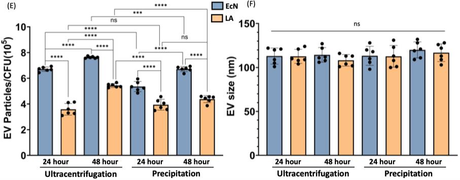
Figure 2. Quantitative comparison of probiotic EV isolation methods. (Sawant, et al., 2025)
Case Study 2: Systematic Optimization of Fungal EV Isolation by Combined Ultracentrifugation and Size Exclusion Chromatography
A comparative study established an optimized isolation system for Fusarium oxysporum extracellular vesicles by evaluating three primary methods (ultracentrifugation, ultrafiltration, PEG precipitation) paired with two secondary methods (size exclusion chromatography, aqueous two-phase system). Solid culture combined with ultracentrifugation-SEC yielded the highest purity fractions, exhibiting typical cup-shaped bilayer membrane structures with a mode diameter of 200.60 nm and mean diameter of 253.50 nm. This combination achieved a particle concentration of 2.04 × 10¹⁰ particles/mL and a particle-to-protein ratio of 1.09 × 10⁸ particles/μg, significantly outperforming ultrafiltration-SEC, PEG-SEC, and ATPS-based alternatives. The study further identified nine candidate fungal EV marker proteins through proteomic analysis, including Hsp70, SNARE proteins, and Rho GTPases, providing a methodological foundation for standardized fungal EV isolation.
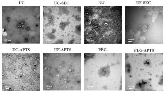
Figure 3. TEM characterization of fungal EV morphology across isolation methods. (Lou, et al., 2025)
FAQs
Q: What is the difference between microbial exosomes and mammalian exosomes?
A: Microbial extracellular vesicles (mEVs) and mammalian exosomes share an overlapping size range, with mammalian exosomes typically 30–150 nm and microbial EVs typically 20–300 nm (fungal EVs occasionally larger), but differ significantly in composition and biogenesis. mEVs from Gram-negative bacteria often contain outer membrane vesicle (OMV) components and lipopolysaccharide (LPS), while Gram-positive-derived EVs may carry lipoteichoic acid (LTA) and peptidoglycan fragments. Yeast EVs present cell wall polysaccharide surface signatures. These microbial-specific components create distinct isolation challenges–including endotoxin control and debris removal—that require specialized workflows beyond standard mammalian exosome protocols.
Q: Which purification method is best for functional studies?
A: For functional cell-based assays, we recommend size exclusion chromatography (SEC) or density gradient ultracentrifugation. SEC effectively removes free proteins and nucleic acids while maintaining EV structural integrity and biological activity. Density gradient ultracentrifugation provides the highest purity, minimizing lipoprotein and aggregate contamination that could confound functional readouts. Both methods yield preparations with low endotoxin levels suitable for immune cell assays, barrier repair studies, and uptake experiments.
Q: Do you support low-endotoxin EV preparation?
A: Yes. We offer optimized low-endotoxin protocols specifically designed for microbial EVs. Through a combination of selective precipitation, affinity-based LPS/LTA removal, and stringent buffer exchange, we can reduce endotoxin levels to thresholds compatible with sensitive in vitro and in vivo applications. Endotoxin quantification via LAL assay is included in our standard QC package.
Q: Can you handle large-scale production?
A: Yes. Our tangential flow filtration (TFF) platform is designed for scalable concentration and purification of large fermentation volumes. We routinely process 500 mL to 100 L+ batches and can develop continuous-flow protocols for pilot-scale manufacturing. This scalability supports progression from laboratory research through preclinical process development without changing the fundamental purification chemistry.
Q: Do you offer customized isolation protocols?
A: Absolutely. We recognize that microbial strains vary significantly in secretion profiles, culture viscosity, and EV surface properties. Our scientists work directly with clients to design customized protocols based on strain identity, culture volume, target particle characteristics, and downstream application—whether that is basic characterization, multi-omics analysis, or drug delivery formulation.
Q: What quality control data will I receive?
A: Standard deliverables include nanoparticle tracking analysis (NTA) for concentration and size distribution, transmission electron microscopy (TEM) images, BCA protein quantification, and LAL endotoxin assay results. Optional advanced QC packages include Western blot for EV marker proteins (e.g., OMPs for bacterial EVs), RNA cargo analysis, proteomics profiling, zeta potential measurement, and stability monitoring under accelerated storage conditions.
References
- Sawant, H., et al. (2025). A simplified method for the isolation of extracellular vesicles from probiotic bacteria and their characterization. International Journal of Molecular Sciences, 26(3), 1058.
- Lou, J., et al. (2025). Establishment of an Isolation System for Extracellular Vesicles of Fusarium oxysporum and Its Proteomic Analysis. Journal of Fungi, 11(12), 884.


