Exosome Functional Validation & Mechanism of Action Studies
OverviewServicesSamplesAdvantagesApplicationsCase StudyFAQs
Overview
At Creative BioMart Microbe, we provide comprehensive functional validation and mechanism-of-action (MoA) studies tailored specifically for microbial extracellular vesicles (mEVs), including bacterial outer membrane vesicles (OMVs), probiotic-derived exosomes, fungal extracellular vesicles, and phage-derived vesicles. Our platform bridges analytical characterization and quality analytics to biological relevance, delivering a complete workflow from cellular uptake quantification and immunomodulatory potency assessment to signaling pathway validation and in vivo efficacy demonstration. Unlike generic mammalian EV service providers, we have optimized every functional assay for microbial vesicle biology—accounting for unique membrane compositions, immunogenic cargo, and host-microbe interaction dynamics.
Our mechanism-driven approach integrates cell-based bioactivity assays, high-resolution imaging, multi-omics profiling, and disease-relevant in vitro and in vivo models. Each project is designed with rigorous positive and negative controls, benchmarked against known inducers where applicable, and documented to support CMC, lot-release, and IND-enabling strategies. Whether you are validating engineered exosomes for drug delivery, screening probiotic exosomes for gut barrier repair, or mapping the immunomodulatory mechanism of OMV-based vaccine adjuvants, our platform provides the functional evidence required for translational decision-making.

Figure 1. Schematic overview of the integrated functional validation and mechanism-of-action studies platform for microbial extracellular vesicles, spanning cellular uptake, immunomodulatory potency, barrier integrity, multi-omics pathway validation, and in vivo efficacy assessment.
Creative BioMart Microbe offers end-to-end microbial exosome functional validation, from project consultation to regulatory-ready data delivery. Learn more about our complete Microbial Exosome Services portfolio. Contact us for a custom quote and project consultation.
Services
Service Workflow

Service Details
Cellular Uptake, Trafficking & Functional Cargo Delivery
This service quantifies microbial exosome internalization and cargo delivery. We use fluorescent labeling, confocal microscopy, flow cytometry, and high-content imaging at ~200–250 nm lateral resolution (diffraction limit; sub-diffraction resolution available via STED or SIM upon request). Kinetics are tracked from 0.5 to 24 hours. Organelle co-localization maps trafficking to lysosomes, ER, or mitochondria. Reporter assays confirm cargo delivery. Deliverables include uptake data and reports for macrophages, dendritic cells, epithelial and tumor lines.
Bioactivity Profiling & Mechanism-Driven Potency Assessment
This service delivers CQA-aligned potency data across three pillars: cellular interaction, biological potency, and cargo delivery. We quantify cytokine panels, macrophage polarization, and T-cell activation. NF-κB activation is monitored by luciferase reporter assays and p65 phosphorylation; NLRP3 inflammasome activation is monitored by ASC speck formation, caspase-1 p20 cleavage, and IL-1β / IL-18 secretion, with optional NLRP3 oligomerization assessment. Readouts include angiogenesis, enzymatic activity, viability, proliferation, apoptosis, and ROS detection.
Comparative Strain Screening & Batch Consistency Analysis
This service compares exosome functional profiles across microbial strains, engineered variants, or manufacturing batches. Standardized potency assays identify high-function sources and quantify inter-batch variability. We correlate physicochemical attributes to biological performance, generating statistical comparison matrices with CV analysis and CQA trending. Clients receive ranked strain reports with consistency scores. This supports strain selection, process optimization, and scaling to application-grade manufacturing.
In Vitro Functional Disease Models & Barrier Systems
This service validates microbial exosome function in physiologically relevant disease contexts. We specialize in intestinal epithelial barrier systems with TEER monitoring, tight junction analysis, and FITC-dextran permeability assays. Co-culture systems integrate immune cells with epithelial layers to model mucosal inflammation, including gut-on-chip platforms. Additional models include wound healing, tumor co-cultures, and LPS-induced inflammation. Each model includes positive controls.
In Vivo Efficacy, Safety & Biodistribution Studies
This service translates in vitro findings into preclinical evidence. We execute efficacy studies using DSS colitis, skin inflammation, tumor, wound healing, and dysbiosis models. Endpoints include histopathology, cytokine profiling, immune infiltration, and IVIS biodistribution. Safety evaluation covers acute toxicity, tolerability, and immunogenicity. Validated functional profiles guide tier selection for Application-Grade Manufacturing and Exosome Formulation & Stability development.
Service Specifications & QC Standards
 Functional Assay Capability
- Our platform covers three integrated functional pillars: Interaction (cellular uptake, trafficking, and cargo delivery), Potency (immunomodulation, barrier repair, angiogenesis, and enzymatic activity), and Drug Delivery Validation (reporter gene functional transfer, release kinetics correlation, and target-cell specificity). Each method is validated with positive and negative controls per batch, and instrument calibration is performed with traceable standards. Assays are designed in alignment with MISEV2023 guidelines and can be adapted to GxP-compliant formats for regulatory submissions.
Typical Data Range
- Uptake detection sensitivity: >90% by flow cytometry
- Quantifiable internalization time window: 0.5–24 hours
- Multi-channel imaging resolution: ~200–250 nm lateral resolution (diffraction limit; sub-diffraction resolution available via STED or SIM upon request)
- TEER monitoring precision: ±5%
- Barrier integrity analysis duration: 24–120 hours
- Permeability tracer sensitivity: 4 kDa FITC-dextran
- Cytokine multiplex detection limit: <1 pg/mL
- Potency assay inter-batch CV: <20%
Turnaround Time
| Project Type |
Timeline |
| Cellular uptake & trafficking assay |
1–2 weeks |
| Cytokine profiling & immunomodulation |
1–2 weeks |
| Cell functional assays (proliferation/migration) |
2–3 weeks |
| Barrier integrity & co-culture studies |
2–4 weeks |
| Comparative strain/batch screening |
3–4 weeks |
| Multi-omics + pathway validation |
4–8 weeks |
| In vivo pilot studies |
6–10 weeks |
| Full in vivo efficacy studies |
8–12 weeks |
| Integrated mechanism studies |
8–16 weeks |
Timeline may vary based on sample complexity, model availability, and assay customization.
Deliverables
- Experimental protocols and SOP summaries
- Raw data files (imaging datasets, flow cytometry FCS files, MS raw data, sequencing FASTQ)
- Processed analytical reports with statistical analysis and publication-ready charts
- Certificate of Analysis (CoA) per batch
- Methodology summary and instrument calibration records
- Bioinformatics analysis report for multi-omics projects (pathway enrichment, regulatory network visualization, and mechanism interpretation)
- Optional CQA documentation package for IND-enabling studies
Quality Control
- Batch-level instrument calibration with certified positive and negative controls
- Inter-batch consistency assessment (functional endpoint CV <20%)
- MISEV2023 compliance checklist for all functional assays
- Contaminant screening for cell debris, protein aggregates, and residual culture media
- Endotoxin monitoring for all in vivo-grade samples
- Optional GxP-aligned assay validation and CQA trending analysis for lot-release documentation
Sample Requirements
| Required Information |
Optional Information |
Not Accepted |
- Sample type (purified microbial exosomes, engineered formulations, fermentation supernatants, lyophilized exosomes, conditioned media)
- Estimated particle concentration or total protein yield
- Strain background and culture conditions
- Sample volume (≥500 μL recommended for in vitro; ≥1 mL for multi-omics)
- Endotoxin level (for in vivo studies)
|
- Prior isolation method (TFF, SEC, ultrafiltration, density gradient, or other)
- Target application (research, CMC, regulatory filing, publication)
- Special assay requests (fluorescent labeling, reporter gene loading, in vivo imaging compatibility)
- Control sample requirements (untreated, vehicle-only, or competitor benchmark)
- Desired functional endpoints or mechanism hypotheses
|
- Samples subjected to more than three freeze-thaw cycles
- Samples with unidentified strain origin or undocumented culture conditions
- Severely degraded, contaminated, or detergent-heavy preparations
- Samples preserved with fixatives, antimicrobial agents, or non-sterile buffers
- Samples shipped at inadequate temperature or with compromised cold-chain documentation
|
Recommended Sample Quantity by Application:
| Application |
Recommended Amount |
| Cell uptake assays |
≥100 μg total protein or 1×10⁹ particles |
| Cytokine & immunomodulation assays |
≥200 μg total protein |
| Barrier integrity studies |
≥300 μg total protein |
| Multi-omics studies |
≥500 μg total protein or 2×10¹⁰ particles |
| Comparative strain screening |
≥200 μg per strain (minimum 3 strains) |
| In vivo pilot studies |
≥1 mg total protein |
| Full in vivo efficacy studies |
≥2–5 mg total protein |
| Biodistribution studies |
≥1 mg labeled exosomes |
Storage & Shipping: Ship frozen at –80°C on dry ice. Store at –80°C upon receipt. Avoid repeated thawing. Recommended buffer: sterile PBS, pH 7.4, endotoxin-free.
Our Advantages
- Microbial EV Functional Specialization: Deep expertise in bacterial OMVs, probiotic and fungal EVs. Assays optimized for microbial membranes, not generic mammalian templates.
- Uptake-to-Pathway Closed-Loop Validation: Integrated workflow from uptake quantification to pathway activation. Enables true mechanism-of-action elucidation, not isolated endpoint readouts.
- Mechanism-Driven Potency Framework: Bioactivity assays structured as potency tests with positive controls and benchmark standards. Directly supports CQA documentation and lot-release strategies.
- Microbial-Specific Endotoxin Control: Dedicated endotoxin monitoring and removal validation for all in vivo-grade samples, ensuring safety profiles aligned with OMV and probiotic EV therapeutic applications.
- DoE-Driven Assay Optimization: Design-of-Experiments approach for multi-parameter assay refinement, maximizing statistical power while minimizing sample consumption for high-value mEV preparations.
- Rapid Turnaround with Milestone Transparency: Standard in vitro packages in 1–3 weeks, in vivo pilots in 6–10 weeks, with QC checkpoint updates and interim data review at every stage.
Applications

Biomarker Discovery & Mechanistic Insight Generation
Multi-omics cargo profiling combined with functional readouts identifies disease-associated signatures and maps mechanistic pathways of mEV-host modulation.

Therapeutic EV Process Development & CMC Support
Functional CQA monitoring, potency validation, and regulatory-ready data packages accelerate mEV transition from preclinical validation to clinical manufacturing.

Probiotic, Cosmetic & Food-Grade Functional Validation
Potency validation for probiotic gut barrier repair, skin cell response for cosmetics, and GI stability for food-grade mEV ingredients.

Vaccine Adjuvant & Drug Delivery Carrier Mechanism Studies
OMV adjuvant qualification, carrier uptake validation, cargo delivery confirmation, and target-cell mechanism mapping for microbiome therapeutics.
Case Study
Case Study 1: Commensal Bacterial EVs Modulate Macrophage Polarization via STING Pathway Inhibition
The researchers demonstrated that extracellular vesicles from Streptococcus hyointestinalis—isolated from cold-tolerant Min pigs—alleviate DSS-induced colitis by targeting the STING-IRF3-TBK1 axis. In wild-type mice, EV gavage suppressed STING overactivation, reduced M1 macrophage markers (CD86, iNOS), and promoted M2 polarization (CD206, Arg1), as validated by immunohistochemistry, Western blot, and flow cytometry. Critically, these protective effects were abolished in Sting-/- mice, confirming STING-dependency. Mechanistically, fluorescently labeled EVs localized to macrophage nuclei and bound STING protein competitively with c-di-AMP. H151 inhibitor assays further verified that EV-mediated M2 polarization requires intact STING signaling. This study establishes a microbial EV–STING–macrophage regulatory axis and demonstrates rigorous mechanism-of-action validation through genetic knockout, pharmacological inhibition, and multi-modal functional readouts.
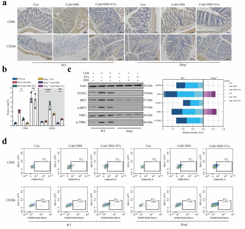
Figure 2. Multi-modal functional validation of S. hyointestinalis EV mechanism of action. (Sun, et al., 2026)
Case Study 2: Bacterial EV-Mediated Immune-Indirect Blood–Brain Barrier Disruption Validated by Organ-on-Chip
This study demonstrated that Escherichia coli–derived bacterial extracellular vesicles (BEVs) indirectly compromise blood–brain barrier (BBB) integrity through immune-mediated mechanisms rather than direct endothelial toxicity. Using a microphysiological system with isogenic hiPSC-derived brain microvascular endothelial cells and pericytes separated by a 100 nm porous silicon nitride membrane, direct BEV or LPS exposure upregulated ICAM-1 without disrupting tight junctions or permeability. Critically, conditioned medium from BEV-stimulated THP-1 macrophages significantly increased TNF-α and IL-1β secretion, caused Claudin-5 gap formation, and elevated lucifer yellow permeability. This study establishes the necessity of incorporating immune components in BEV mechanism-of-action studies and validates a multi-cellular organ-on-chip approach for dissecting indirect EV-mediated barrier disruption.
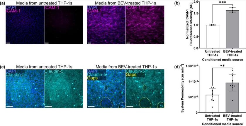
Figure 3. Functional validation of BEV-mediated indirect BBB disruption. (Widom, et al., 2026)
FAQs
Q: What types of microbial exosomes can be functionally evaluated?
A: We support exosomes and extracellular vesicles derived from Gram-negative and Gram-positive bacteria, probiotics, fungi, yeast, phage-derived vesicles, and engineered microbial strains.
Q: How much sample is required for uptake and immunomodulation assays?
A: Cellular uptake assays require ≥100 μg total protein or 1×10⁹ particles. Cytokine profiling and immunomodulation assays require ≥200 μg. In vivo studies require ≥1–5 mg depending on model complexity and dosing regimen.
Q: Can you design customized functional assays for novel mechanisms?
A: Yes. We provide fully customized assay development based on your target application, disease model, or mechanism hypothesis. Custom endpoints, cell types, and pathway reporters are available upon consultation.
Q: Do you provide in vivo efficacy and biodistribution studies?
A: Yes. We offer pilot studies (5–10 animals per group) and full efficacy studies (10–20 animals per group) using colitis, skin inflammation, tumor, wound healing, and microbiome-associated disease models with IVIS biodistribution and histopathological endpoints.
Q: What signaling pathways can you validate?
A: We routinely validate NF-κB, MAPK, PI3K/AKT, JAK/STAT, Wnt/β-catenin, TLR signaling, and autophagy pathways. Custom pathway validation using reporter assays, inhibitor studies, siRNA knockdown, or CRISPR-based target editing is available upon request.
Q: Can engineered exosomes be validated for functional performance?
A: Yes. We evaluate drug-loaded, surface-modified, reporter-gene-loaded, and genetically engineered exosomes for uptake efficiency, cargo functional delivery, target-cell specificity, and mechanism of action.
Q: Do you support publication and regulatory documentation?
A: Yes. We provide publication-ready datasets, high-resolution images, statistical analysis, bioinformatics reports, and optional CQA documentation packages suitable for IND-enabling studies, cosmetic raw material registration, and food-grade safety filings.
Q: How should functional validation samples be shipped?
A: Ship frozen at –80°C on dry ice. Avoid repeated freeze-thaw cycles (maximum three recommended). Use sterile, endotoxin-free PBS. We provide detailed shipping guidelines and perform incoming quality inspection upon receipt.
References:
- Sun, Z., et al. (2026). Microbial extracellular vesicles from min pigs remodel macrophage polarization via STING to sustain intestinal immune homeostasis. Gut Microbes, 18(1), 2620126.
- Widom, L. P., et al. (2026). Bacterial extracellular vesicles indirectly destabilize a human stem cell-derived blood-brain barrier on-chip through pro-inflammatory stimulation of immune cells. bioRxiv, 2026-01.
Related Services:


